Feature Story

Canadian 3D technology is solving problems of the heart
July 7, 2016
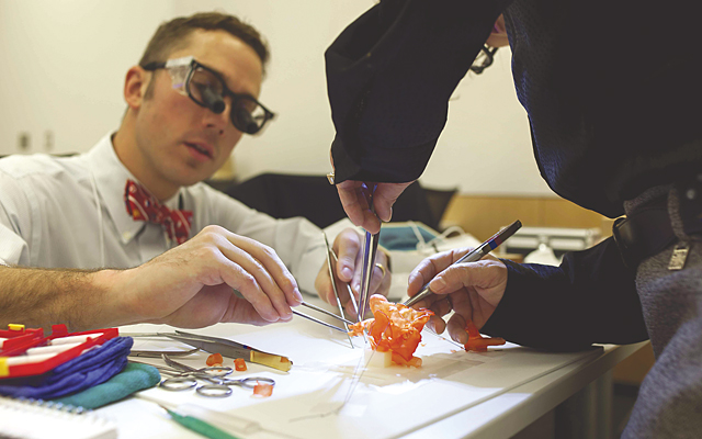
A tiny polymer heart the size of an adult ear, with an even tinier patch stitched on to it, provides deep insights to the surgeon preparing to operate at Toronto’s Hospital for Sick Children. But this is no ordinary model of a typical baby’s heart.
It’s an exact replica of a particular infant’s heart – including the patient’s own complex congenital defect. The intricate model enables a difficult operation to be rehearsed, and reduces the risk well before the child’s scheduled surgery.
Nearby, in a chemical engineering lab at the University of Toronto, a team is growing living heart cells, not in a petri dish, but on a unique polymer scaffold that effectively tricks the cells into thinking they are growing as part of a living heart, and so they start beating as if they were. The tissue eventually may be used as replacement tissue, but currently is highly valued for use in testing new heart drug compounds.
These are two of the latest Canadian-driven developments that use 3D-printing technology as an aid in solving heart-related medical issues.
At The Hospital for Sick Children, Dr. Glen Van Arsdell, cardiovascular head and Dr. Shi-Joon Yoo, a cardiac radiologist who leads the hospital’s Division of Cardiac Imaging, launched a program that scans the hearts of very young patients with congenital defects and creates 3D-printed replicas on which surgeons can practice incisions and sewing patches or performing other operations.
Developing the model hearts, made of a polymer resin, starts with CT, MRI and ultrasound scans with sectional imaging, so they can view all the multiple structures overlapping each other through projectional images.
“After the CT and ultrasound, we do sectional imaging so we can see the inside of the body in detail. We can decide to do 3D reconstructive, or volume rendering. But it’s on the computer screen,” says Dr. Yoo. With a polysial replica, it’s a different story: “When you have the complex geometry in your hand, it’s easier to understand,” he adds.
Communication between the radiologist and the surgeon is improved dramatically as well. “It can take 15 to 20 minutes to explain what is in the images, and with these replicas we don’t have to explain,” says Dr. Yoo.
The ability to use a replica heart for surgical training is particularly useful when dealing with tiny infant hearts. For example, explains Dr. Yoo, “The Norwood Procedure for hypo-plastic left heart syndrome. The geometry is complex, and you’re dealing with a very tiny heart. Any surgeon has difficulty learning how to do it. So the training is incredibly important.”
Before, says Dr. Yoo, a lot of patients were at risk when surgeons needed to learn this. Now, with practice, they can significantly reduce the risk. Also, he adds, “If the surgeon understands the exact anatomy (he/she) doesn’t have to spend time exploring during the surgery.” Even assistants can gain a better understanding of what’s going on, what the operation is doing for the patient by studying the replica hearts. “All this together leads to better outcomes,” he adds.
Doctors graduating from medical school can wait up to 15 years before even getting to participate in surgeries involving certain types of infant heart defects. A big reason for that is the lack of hearts to practice on, that have to come either from animals or donors who have died. In an educational setting, notes Dr. Yoo, no one has to die to provide a heart. “We can show the full spectrum of congenital heart defects and we can send the hearts anywhere in the world.”
Asked about similar efforts involving 3D-printed hearts, Dr. Yoo, who has a reputation as the foremost expert in this area, said “A lot try to copy it, but it is really not copy-able. You need to invest a lot of time. What is in the images is not 100 per cent of it – you need an intellectual component added and also years of practice.”
The cost of developing the replicas can also be prohibitive. From scratch it would cost about $1,500 for each piece, but when considered as a business the cost would be about $5,000 each,” he explains.
Clearly 3D- printing of replica hearts provides radiologists and surgeons with deep insights that assist in achieving better outcomes. Asked whether there might be a time when a 3D-printed heart could replace a human heart, Dr. Yoo said “It’s not at the applicable stage. Eventually I think so.”
The team led by University of Toronto tissue engineer Milica Radisic, who was named to the Royal Society of Canada’s new college and is a fellow of the American Institute for Medical and Biological Engineering (AIMBE), is using 3D technology in a different way. They are using 3D-printed scaffolds as a support structure for growing swatches of living heart tissue.
“We build living heart tissue and liver tissue in the lab,” says Professor Radisic, who, along with her team has been working on this achievement for the past 10 years.
While most other efforts to build heart tissue are conducted in a petri dish, using a 3D-printed scaffold to encourage growth is unique. “A petri dish is just a petri dish. The cells sit at the bottom. It’s pretty rigid,” explains Dr. Radisic.
The scaffold, made of polymers and hydrogel, provides a more natural growth environment. The 3D polymer scaffolds called AngioChips, or person-on-a-chip technology, are built by Dr. Radisic’s students in a clean room – similar to the environment used for building computer chips.
“We didn’t want to harvest blood vessels,” says Dr. Radisic, adding that they also wanted a stable structure to use. “We created branch and blood vessels out of polymer,” she says. AngioChip behaves like vasculature, and around it is a lattice for other cells to attach and grow. The original design for the scaffold was enhanced significantly, with pores that blood vessels can grow through, by Boyang Zhang, a PhD student in Radisic’s lab.
Asked about the source for the growing heart cells, Dr. Radisic said, “One problem with the heart, and using induced Pluripotent Stem cells (iPS cells) obtained from an adult to grow heart tissue, is we cannot take cells out of your heart. They don’t divide after birth – well, they don’t divide fast enough. They divide very, very slowly and no one’s figured out how to make them divide faster. That’s why we have to use stem cells.”
“We are reprogramming the stem cells we obtain from the foreskin of newborn babies at Mt. Sinai Hospital,” explains Dr. Radisic, adding that the only difference beyond that to make the heart cells, in addition to using the 3D scaffolds, is that they also apply hydrogels and electric stimulation to the stem cells.
