Feature Story
Rethinking the practice of pathology by using AI
September 30, 2020
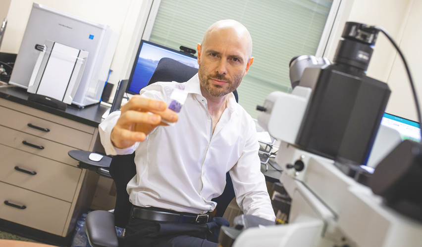
HAMILTON, Ont. – Dr. Clinton Campbell envisions a future where pathologists work as information specialists in sophisticated computer labs with the world’s most current, relevant data at their fingertips. Instead of diagnosing disease one slide at a time under a microscope, there would be thousands of images to compare online.
Dr. Campbell is working to make that vision possible by harnessing a type of artificial intelligence (AI) known as machine learning.
The first steps toward this high-tech future are taking place at Hamilton Health Sciences (HHS) right now, thanks to an innovative pilot project exploring digital pathology and AI. It’s among the first projects of its kind in Canada, and could help modernize the way pathology is conducted around the world.
“Pathologists currently use an old-fashioned light microscope to view sections of human tissue, which is a 300-year-old technology,” said Campbell, one of about 100 pathologists in Canada who specializes exclusively in blood disorders, like leukemia. Campbell is leading an HHS study into digital pathology and AI. He is supported by Drs. Catherine Ross and Monalisa Sur for this pilot.
Pathologists study tissue samples of disease taken from patients during surgery, or in the case of blood, during a procedure known as a bone marrow biopsy.
Samples are placed on glass slides for examination under a microscope. Pathologists then look at minute features of cells and tissue and extract information to better understand the disease. This, in turn, may lead to a diagnosis and the best treatment options for patients.
“The system we have currently works well,” said Dr. Campbell. “But we can’t share images of human tissues in real-time with colleagues or do `virtual peer review’ where multiple pathologists can examine slides together at the same time.”
Currently, when a pathologist at another hospital wants Dr. Campbell to view a set of slides and offer insights, the slides are shipped to him in thick cardboard folders.
“This is an inefficient process that leads to delays in diagnosis. Imagine how much better it would be if we could share these slides digitally in real-time,” said Dr. Campbell. “It would allow for wider, faster consultation. And if you add AI into the mix, you’ve also got access to a vast amount of knowledge that’s sorted through machine learning to prioritize the very best information to help with your case.”
Machine learning, in the most basic sense, is a type of AI that can automatically learn patterns from data, like pathology images, and then use this knowledge to make useful predictions about the world. This means faster, better reports because machine learning will help extract the most important information, and then compare this to tens, hundreds or even thousands of other previously diagnosed samples instantly.
“It’s kind of like having Google for pathology, with a team of virtual pathologists on standby in the cloud 24/7,” he says. “This could transform the way diagnostic medicine is done.”
HHS is conducting a one-year pilot project on digital pathology in partnership with Huron Digital Pathology in St. Jacobs, Ont.
Huron’s AI-based image search platform connects pathologists to the expertise of their colleagues worldwide, increasing the speed and effectiveness of patient care.
HHS is using a scanner provided by Huron to collect and catalogue digital slide images. The project started in May and is based at HHS Juravinski Hospital and Cancer Centre. The goal is to collect 3,000 digital images from blood and bone marrow pathology slides.
The partnership between HHS and Huron was made possible through the Ontario Bioscience Innovation Organization (OBIO), a not-for-profit organization dedicated to advancing health technology innovation and commercialization. The OBIO recently announced the launch of the first eight Early Adopter Health Network (EAHN) innovation projects, including the Huron-HHS partnership.
So why has it taken so long for pathology to dip its toes into the digital pool? A major hurdle has been image file size and processing. Hamid Tizhoosh heads the Laboratory for Knowledge Inference in Medical Image Analysis (KIMIA Lab) at the University of Waterloo. He helped Huron develop its technology and has been working with Huron and Dr. Campbell to devise a system that loads images faster and more efficiently.
They are doing this by collecting only the small sections of pathology specimen images that relate directly to the disease, rather than trying to load the entire image. Images are then processed with AI tools to make a powerful image search engine.
“Huron and the KIMIA Lab piloted this groundbreaking technology, and we’re now expanding this into hematology with the hope of improving it and applying it to blood disorders like leukemia, lymphoma and myeloma,” said Dr. Campbell.
Lise Diebel is a Public Relations Specialist at Hamilton Health Sciences.